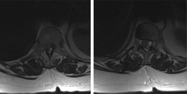
Fig. (2)
Magnetic resonance tomography: Axial layer in T2- sequence, presenting the high signal intensity of the intradural haematoma at the levels of the 12
th
thoracic and the 1
st
lumbar vertebra.