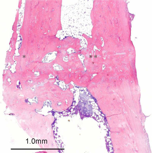
Fig. (9) Histological longitudinal section through the rat femur at
16 weeks postoperatively (magnification ×40, hematoxylin and
eosin staining): membranous ossification in the subperiosteal
region(*) and the primary fusion from endochondral ossification
which were displayed proliferation of osteoblasts, chondrocytes and
neovascularization (**).