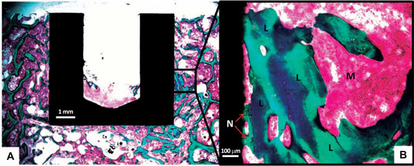
Fig. (4)
(
A
) Picture showing a histological cross section used for Histomorphometry. The close-up picture (
B
) shows Lamellar Bone (L) and New Bone (N) in contact with the implant surface. Marrow is marked (M).