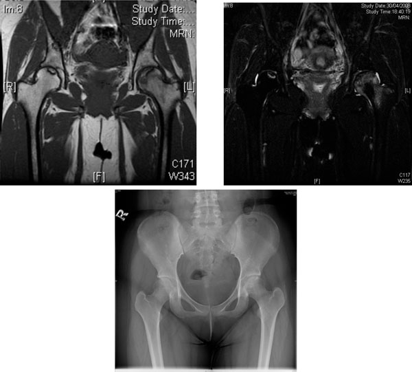
Fig. (1) AVN of the femoral heads in a 19-year-old woman with lupus nephritis treated with long-term glucocorticoid. (a) Coronal T1-
weighted MRI scan. (b) Coronal T2-weighted MRI scan. Heterogeneous area bordered by well defined T1W & T2W hypointense rim was
observed over the superior left femoral head. T1W & T2W hyperintense lesions bordered by T1W hypointense T2W hyperintense rim was
seen at superior right femoral head. Features are suggestive of bilateral AVN, more severe on the left side. (c) X-ray of the hips of the same
patient - increased mixed sclerosis/lucency is seen at subchondral region of superior aspect of left femoral head, the configuration of which is
still preserved (early AVN). The right hip appears unremarkable.