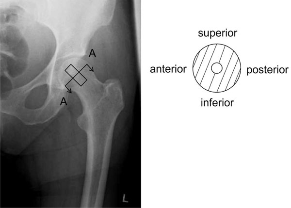
Fig. (1)
Sample orientation in anterior-posterior X-ray (left) and schematically sectional drawing A-A (right). The samples were drilled out of the centre of the human femoral head.