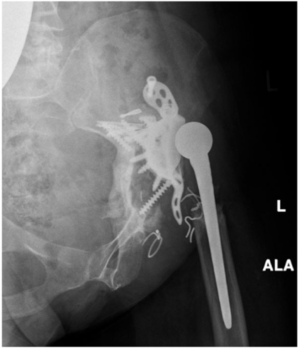
Fig. (4) Compared to standard views (Fig. 2) additional information
with respect to the bony defect situation can be obtained with further
radiological diagnostics. In this case, simple oblique iliac radiograph
shows a highly deficient posterior wall, massive sclerosis of the host
bone and defects at the bottom due to loosening and dislocation of
screws.