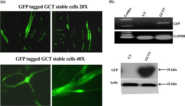
Fig. (1) Subcellular localization of GFP signals in GCT transfected stromal cells. GFP signals were detected by immune-fluorescence.
(b) Semi-quantitative PCR: GFP expression in transfected GCT stromal cells was detected using GFP specific primers and GAPDH was
used as an internal control. UT=untrasnfected cells and GCTT= GCT transfected cells. (c) These results are verified at the protein level by
western blot analysis.