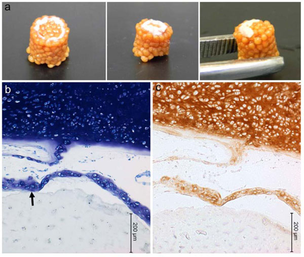
Fig. (5) Porcine mesenchymal stem cells cultured on chitosan-CaP scaffolds for 28 d in chondrogenic medium. a – gross appearance of
constructs formed by Technique #2 (refer to text); b – toluidine blue staining of histological sections (arrow points to scaffold-tissue
interface); c – positive immunostaining for type II collagen as indicated by brown DAB chromagen.