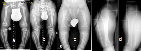
Fig. (1) (a-d) AP radiographs of the lower limbs in achondroplasia (three boys and one girl of 6, 7, 8 and 9 years-old respectively), all
manifested the characteristic radiographic features of achondroplasia; shortness of the tubular long bones, with a relative increase in bony
diameters and densities are apparent. The metaphyses are widened and flared, but the epiphyses are uninvolved. The growth plates are U-shaped
or V-shaped. This is best seen at the distal femur. The long bones, especially the tibiae, are bowed. The pelvis characteristically
appears broad and flat, with squared iliac wings. The ilium appeared broad because the pelvis is formed almost entirely by intramemberanous
ossification, which is undisturbed in achondroplasia. The sciatic notches are small and the acetabuli are horizontal. Genu vara was a constant
skeletal deformity encountered. In addition there was a relative shortening of the tibia compared to the fibular length. This tibial shortening is
typically associated with a relevant ankle joint varus evolving during growth, the varus deformity ranged between 15 to 20°.