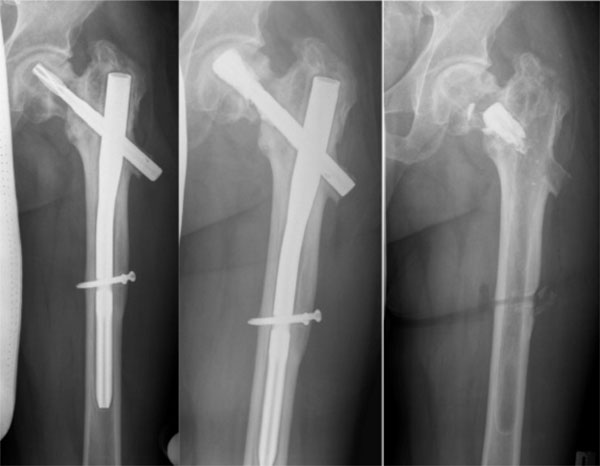
Fig. (5) Patient no. 10 with loosening of the PFNA blade. Good visualization of the bone defect in the left image. In the middle, the
postoperative x-ray after augmentation of the blade is shown. A filling of the defect can be seen. On the right is the x-ray after consolidation
and implant removal after 17 months.