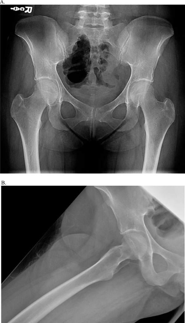
Fig. (1)
AP (A) & lateral (B) views of the pelvis and right hip 3 weeks prior to current ED presentation.