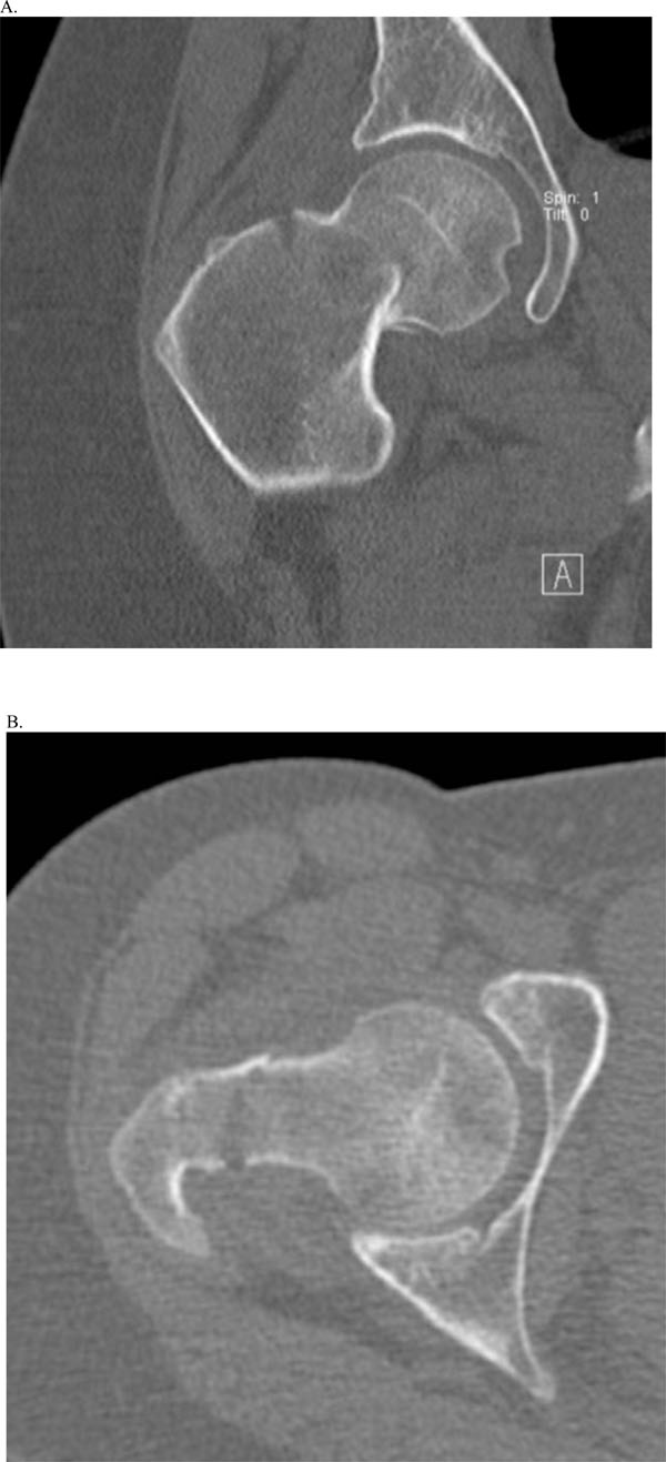
Fig. (4)
Coronal (A) and Axial (B) CT images at the time of presentation.