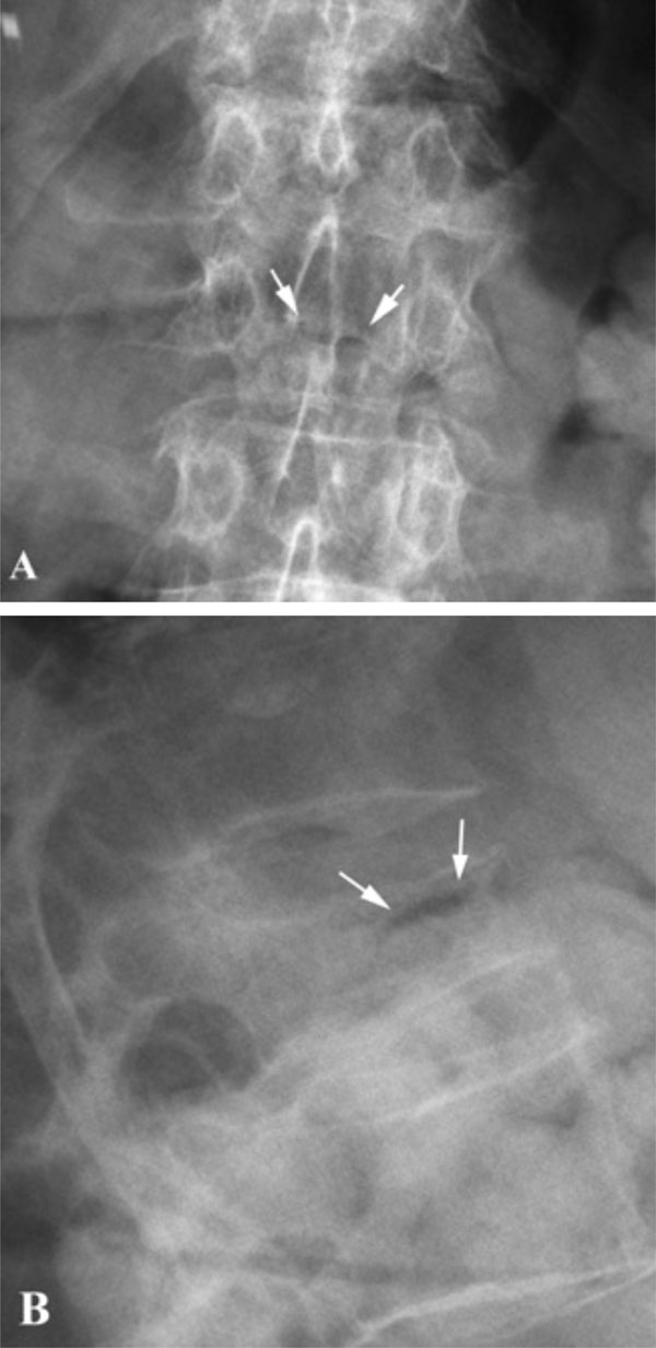
Fig. (1) A 76-year-old female patient, who was under systemic
steroid treatment for rheumatoid arthritis, was presented with
refractory back pain. Anteroposterior (A) conventional radiograph
shows a barely visible horizontal linear IVC in L2 vertebra, which
is more apparent on lateral (B) projection (arrows). The L2 vertebra
is wedge-shaped, while the IVC appears typically at the anterior
third of the vertebral body (B).