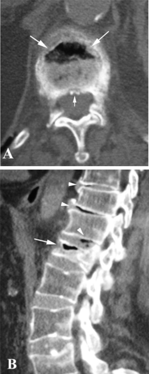
Fig. (2) CT scan of the lumbar spine of the same patient. The
intravertebral vacuum cleft phenomenon is identified in the anterior
third of the L2 vertebral body on both axial (A) and sagittal (B) CT
study of the lumbar spine (large arrows). Minimal retropulsion of
bony fragments is observed (small arrow) (B). Note the
degenerative disc disease and the coexistent intervertebral disc
vacuum phenomena at the levels T11-T12, T12-L1, L1-L2 on the
sagittal CT-scan (arrowheads) (B).