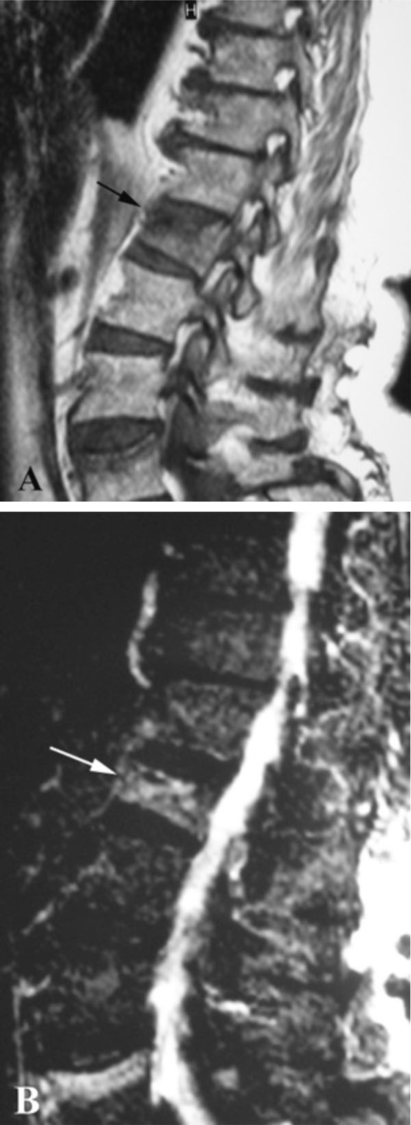
Fig. (3) Sagittal MRI studies of the lumbar spine of the same
patient. The intravertebral vacuum cleft shows low signal intensity
on T1 (A) and high signal intensity on T2-weighted (B) sequences
(arrows). These two different signals represent a fluid collection.