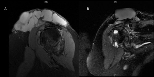
Fig. (2) (a) Sagittal PD (b) Coronal T2 MRI of the right shoulder showing marked degeneration of the glenohumeral joint with a
longstanding chronic full-thickness rotator cuff tear with complete atrophy of the supraspinatus and infraspinatus muscles and a lesser extent
the subscapularis muscle. There is direct bone on bone articulation between the superior and humeral head and the undersurface of the
acromion. Prominent degenerative changes are also noted within the acromioclavicular joint. There is a large multiloculated cystic lesion (9 x
5 cm) relating to the soft tissues overlying the deltoid muscle which has relatively simple features and is consistent with a large ganglion cyst
likely relating to the adjacent markedly degenerated glenohumeral joint and cuff pathology.