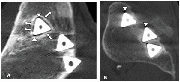
Fig. (6) (A) Sagittal CT scan of the iliac portion of the joint shows a sclerotic margin surrounding the edges of the superior implant. Areas of
“spot-welds” (arrows) noted between the sclerotic margin and implant walls is suggestive of biological fixation. Artifacts are apparent at the
corners of the implant (triangle). (B) The sacral side shows increased bone density adjacent to the implant walls.