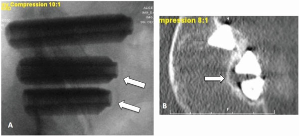
Fig. (7)
A) AP x-ray and B) axial CT image of a non-study patient showing a wide gap between the edge of the implant and the sclerotic margin, indicative of implant loosening. This patient returned for revision surgery.