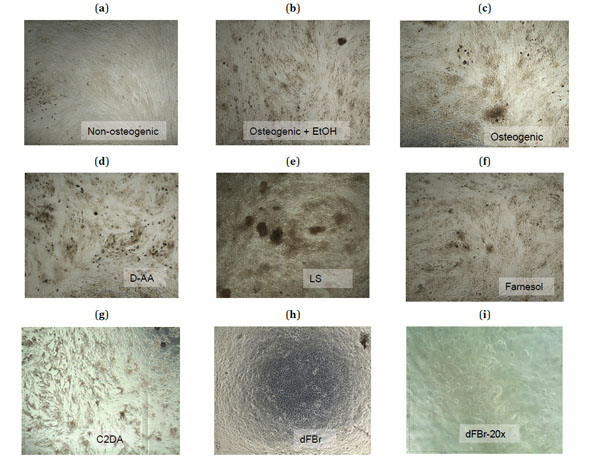
Fig. (2) Microscopic images (4X magnification) taken at day 21 of the control groups (a-c) and the highest sub-toxic concentrations of each
test group (d-h) stained with Alizarin Red-S to show calcium deposits in dark red-brown. A 20X magnification of cells exposed to dFBr (i)
shows the rounded morphology of these cells.