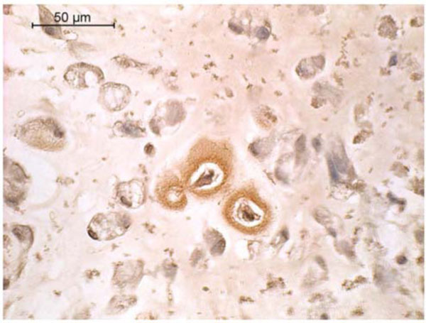
Fig. (5)
Type I collagen immunohistochemistry of canine MSCs embedded in PRP/alginate gel and cultured 4 weeks in defined chondrogenic medium without TGF-β3. Positive staining is indicated by a reddish brown color.