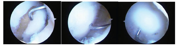
Fig. (8) A series of three arthroscopic images of a medial femoral condylar lesion due to osteochondritis dissecans. The cartilage is intact on
the lateral side but the lesion can be mobilized on the cartilage hinge by the probe (left). After curetting of the fibrous tissue off the bone base
and removing any dead bone, the lesion is reduced anatomically by the probe (central) and then fixed with bioabsorbable pins
arthroscopically (right). One pin can be seen just to the right of the probe and the probe is used to check that the fixation is stable. Although
this is a chronic lesion, the fixation principle is valid for acute lesions.