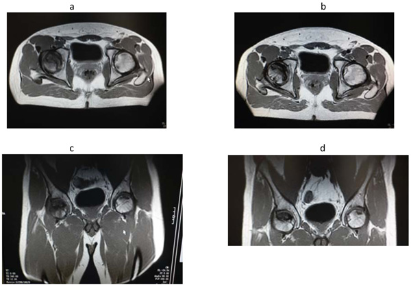
Fig. (1) a – c= MRI images performed at the onset of the disease.b – d= MRI images performed after three month since start of the treatment with intravenous
pamidronate.
Reduction of the perifocal oedema and of the dimensions of the two foci of the head of the left
femur (blue arrows). The necrotic area present in the head of the right femur was unchanged
(orange arrow).