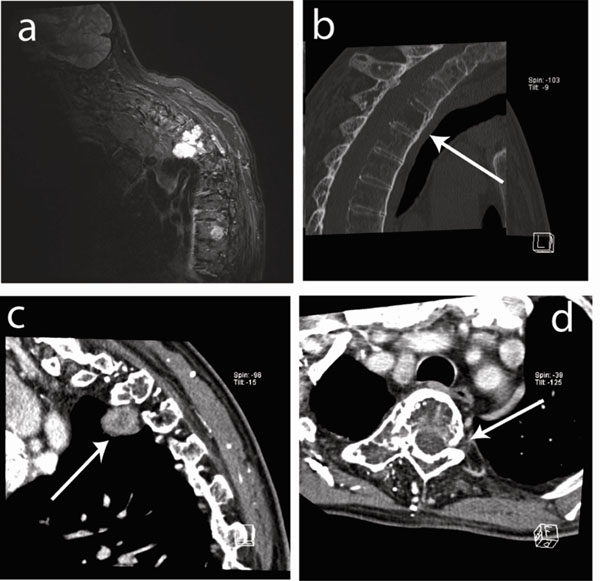
Fig. (2) Imaging of the cervical and dorsal spine. (a) Soft tissue prevertebral mass on the right side of dorsal vertebra 3 (D3) visualized by
STIR MRI. (b) Sagital CT image showing osteolytic lesions (arrow) in vertebrae D3, D4 and D5 (corpus, processus spinosus and
transversus) with diffuse cortical involvement and destruction of the posterior wall. (c) Sagital CT image showing the prevertebral mass on
the right side (3.3x1.7x2.7cm) (arrow). (d) Transverse CT image showing osteolytic destruction (arrow).