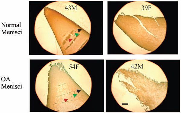
Fig. (4) Immunolocalization of type I collagen. Normal menisci (transverse sections of the inner portion of menisci) showed strong
immunolocalization of type I collagen (top photos). Type I collagen staining in the surface zone (black arrow) of normal menisci was greater
than that in the middle zone (green arrow) and deep zone (red arrow). Type I collagen staining was decreased in OA menisci compared to
normal menisci (compare the bottom photos to the top photos). The reduction of type I collagen staining was much more prominent in the
deep zone (the red arrow) than that in the surface zone (the black arrow). Scale bar: 250 µm.