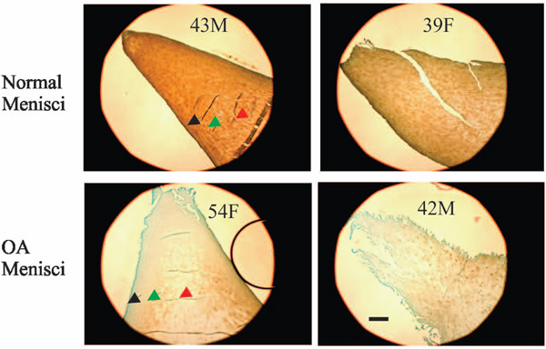
Fig. (5) Immunolocalization of type II collagen. Normal menisci (transverse sections of the inner portion of menisci) showed strong
immunolocalization of type II collagen (top photos). Type II collagen staining in the surface zone (black arrow) of normal menisci was
greater than that in the middle zone (green arrow) and deep zone (red arrow). Type II collagen staining was decreased in OA menisci
compared to normal menisci (compare the bottom photos to the top photos). The reduction of type II collagen staining was prominent in all
the three zones. Scale bar: 250 µm.