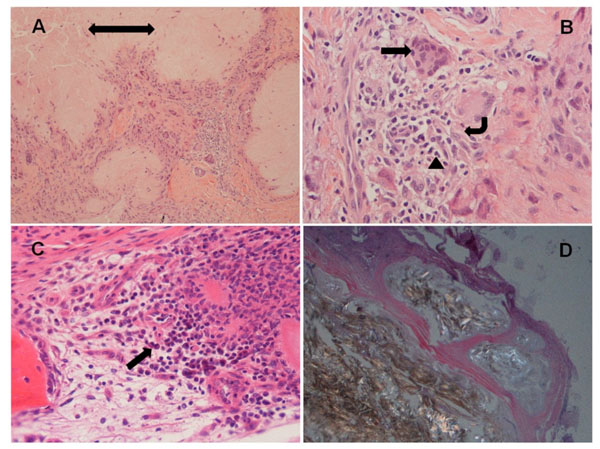
Fig. (1) Examples of the tissue from patients with gout. (A) H&E section shows central amorphous material (double head arrow) rimmed by multinucleated giant cells and inflammatory cells, and surrounded by soft tissue (100X magnification); (B) Multinucleated giant cells (arrow), macrophages (angular arrow) and neutrophils (arrow head) (400X); (C) Amorphous material and inflammatory cells including rare plasma cells (arrow) in bone marrow (400X); (D) Polarized light microscopy demonstrates strongly negatively birefringent needle-shaped crystals and surrounding fibrous tissue (100X). Of note, hand joint synovial soft tissues were showed in (A, B, C); foot joint synovial soft tissue was shown in (D).