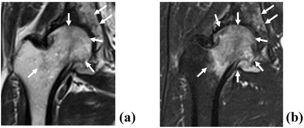
Fig. (4) MRI showing high signal change in the entire femoral head and acetabulum. Broad low intensity signal by T1-weighted image (a)
and high intensity by T2-STIR (arrows) (b) in the right femoral head and acetabulum. Note that there was continuous joint pain when MRI
was performed.