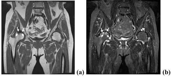
Fig. (6) MRI showing bone cysts and local signal changes in the load-bearing areas of the right femoral head, and no signal change in the
left hip joint. Local low intensity in the femoral head by T1-weighted image (arrow) (a) and high intensity by T2-STIR (arrows) (b) in the
right femoral head.