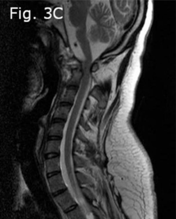
Fig. (3c)
Pre-operative cervical MRI T2-weighted (sagittal view) showing significant reduction of the anterior-posterior diameter of the intra-canal space.