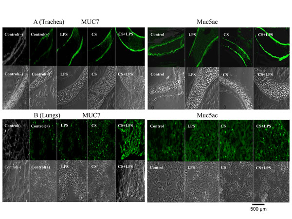
Fig. (3) Effect of CS, LPS and CS/LPS on mucin production in vivo, using MUC7 transgenic mice determined by immunohistochemistry. A representative results from tissues of several mice are shown. MUC7 and Muc5ac in trachea (panel A), and MUC7 and Muc5ac in lungs (panel B). Fluorescent (upper) and phase contrast (lower) pictures are shown for each tissue sample. For MUC7 mucin, Control(-) and Control(+) were tissue samples from trachea or lung samples from the non-treated nontransgenic mice (not expressing human MUC7) and MUC7 transgenic mice (expressing MUC7), respectively. For Muc5ac mucin control, samples from trachea or lung from the non-treated MUC7 transgenic mice (these express Muc5ac) were used. Pictures (200×) were acquired by a digital camera attached to a Nikon inverted fluorescent microscope. Hematoxylin-eosin staining (not shown) indicated the integrity of the tissue.