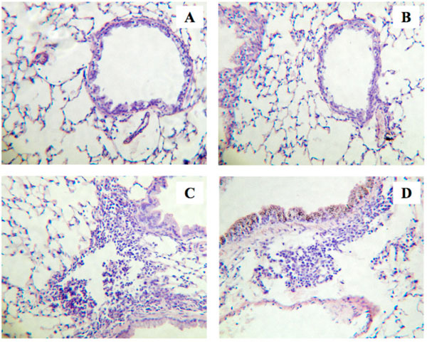
Fig. (1) Histological examinations of lungs from the repeatedly antigen-challenged (Challenged; C and D) and control mice (Control; A and
B). Animals were also received intranasal administration of sphingosine-1-phosphate (S1P; 10-5 M, 20 µL) or its vehicle (1% methanol in
sterile PBS, 20 µL) 30 min prior to each antigen exposure. Four-µm sections of formalin-fixed lung tissues were stained with hematoxylin
and eosin before examination by light microscopy. The photos shown are Vehicle-Control (A), S1P-Control (B), Vehicle-Challenged (C),
and S1P-Challenged groups (D), and are representative of 3 different animals, respectively. Original magnification: x80.