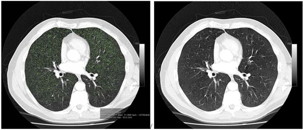
Fig. (1) Chest CT (axial slice) of a 64 year-old male patient.All lung parenchyma with an attenuation value of less than -910 HU is
highlighted in green (left image) using image segmentation software. The volume of the region of interest (green areas) is then calculated.
The corresponding slice is shown on the right prior to application of our technique.