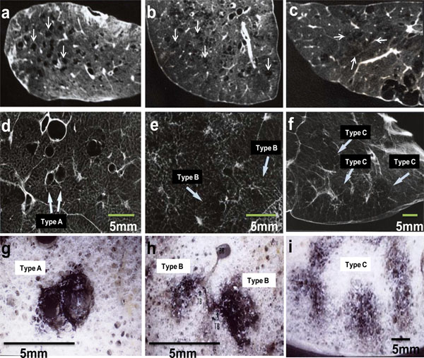
Fig. (2) High-resolution CT, radiograph and stereomicroscopic images of inflated-fixed lungs. High-resolution CT (a, b, c), radiograph
(d, e, f) and stereomicroscopic images (g, h, i) were obtained from specimens of 5 mm-thick slice of inflated-fixed lungs. Type A LAA was
round or oval shape with well-defined border (a, d). The size was less than 5 mm in diameter. Type B LAA was polygonal or irregular shape
with ill-defined border (b, e). The size was less than 5 mm in diameter. Type C LAA was irregular shape with ill-defined border, coalescing
with each other (c, f). The size was 5 mm or over in diameter. Emphysematous lesions are located in a centrilobular site with carbon deposits
(g, h, i). The series of image a-d-g, b-e-h and c-f-i were taken from same specimen, respectively. In addition, d-g, e-h and f-i show one by
one correlations, respectively. TB: terminal bronchiole.