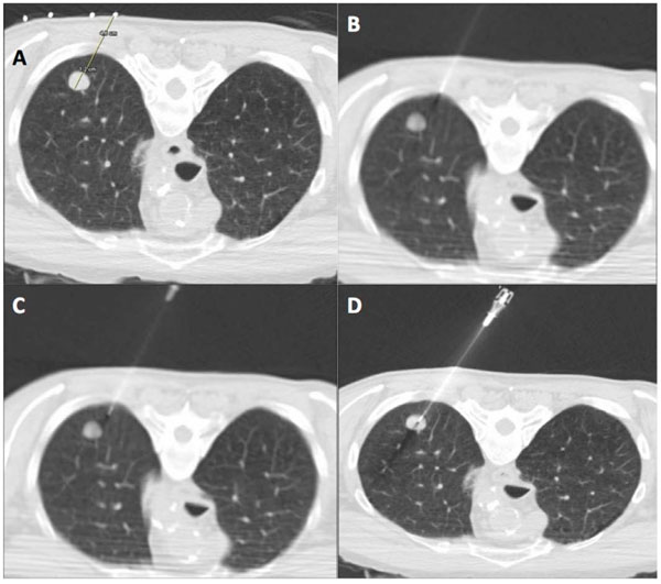
Fig. (2) CT guided percutaneous core biopsy of lung (PCBL). (A) Lesion is localized and access route is planned using thin section CT. The
skin entry site is marked on the patient with indelible ink with use of CT gantry laser light. The distance to the lesion and depth of lesion is
measured. (B) The introducer (19 gauge ultra-thin needle) is carefully aligned in the axial plane with guidance from CT gantry light. The
needle is advanced in small increments through the soft tissue of the chest wall. (C) Once the introducer needle is correctly aligned, a single
deliberate puncture of the pleura is made and the needle is advanced beyond the pleura for at least 2 cm. If needle is misaligned, the
introducer can be repositioned in small increments without exiting the lung. (D) Once satisfactory needle position is confirmed by CT, the
introducer needle is advanced into the edge of the lesion. The inner stylet of the 19 gauge needle is removed and core biopsy performed with
20 gauge automated cutting needle.