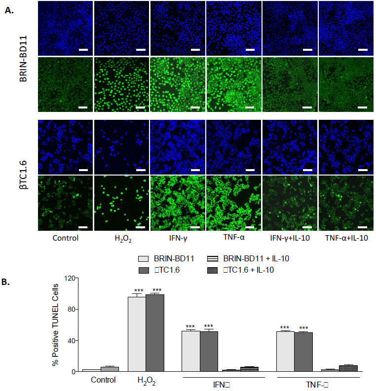
Fig. (7)
Recombinant IL-10 protects against IFN-γ and TNF-α-driven apoptosis.(A) Fluorescent images showing the ability of IL-10 (1 ng/ml) to reduce the % positive TUNEL cells after treatment with a combination of IL-10 and 1 μg/ml of IFN-γ or 1 μg/ml of TNF-α. 1% H2O2 acted as a positive control in these experiments. Blue staining represents DAPI staining of the nuclei while green staining indicates TUNEL positive cells (B) The % positive TUNEL cells were measured by calculating the number of positive TUNEL cells divided by the total number of cells. Data are presented as mean ± standard deviation (SD) with n=3. ***P<0.001 and ****P<0.0001 compared with untreated controls. The scale bars in all images equal 100 µm. IFN-γ, Interferon gamma; TNF-α, tumour necrosis factor alpha; IL-10, Interleukin 10.The viability of BRIN-BD11 (A) and βTC1.6 cells (B) was assessed by colorimetric MTT assay after the addition of 100 ng/ml of anti-IL-10 ± IFN-γ or TNF-α. Data are normalized to untreated controls and presented as mean ± standard deviation (SD). n=3 with all experiments assayed in duplicate. **P<0.01 and ***P<0.001 compared with corresponding non-conditioned media control (i.e., RMPI or DMEM). MSC-CM, Mesenchymal Stem Cell Conditioned Media; IFN-γ interferon gamma, TNF-α tumour necrosis factor alpha.