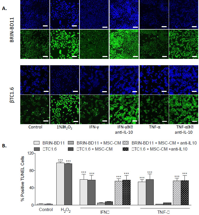
Fig. (9)
Blockage of IL-10 prevents the anti-apoptotic activities of MSC-CM. BRIN-BD11 and βTC1.6 cells were exposed to 1 µg/ml IFN-γ or 100 ng/ml TNF-α, for 24h in the presence or absence of IL-10 depleted MSC-CM (100 ng/ml anti-IL-10 antibody) and the induction of apoptosis assessed by TUNEL assay. (A) Fluorescent images showing the inability of IL-10 depleted MSC-CM to reduce the % of positive TUNEL cells after cytokine challenge. 1% H2O2 acted as a positive control in these experiments. Blue staining represents DAPI staining of the nuclei while green staining indicates TUNEL positive cells (B) The % of positive TUNEL cells was measured by calculating the number of TUNEL positive cells divided by the total number of cells. Data are presented as mean ± standard deviation (SD) with n=3. ***P<0.001 compared with untreated controls. The scale bar in all images equals 100 µm. MSC-CM, Mesenchymal Stem Cell Condition Media; IFN-γ, Interferon gamma; TNF-α, tumour necrosis factor alpha; IL-10, Interleukin 10.