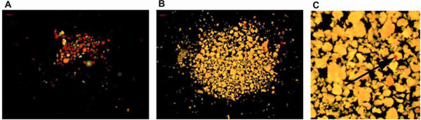
Fig. (1) Dithizone staining of two samples of human pancreatic cell clusters with varying amount of endocrine islets (stained red). The
estimation of endocrine islets in the two samples is (A) 80% and (B) 2% respectively. (C) Enlarged image of the exocrine cell clusters shows
an embedded endocrine islet (arrow). Figure A represents a sample from the endocrine fraction used in the immunohistochemistry study.