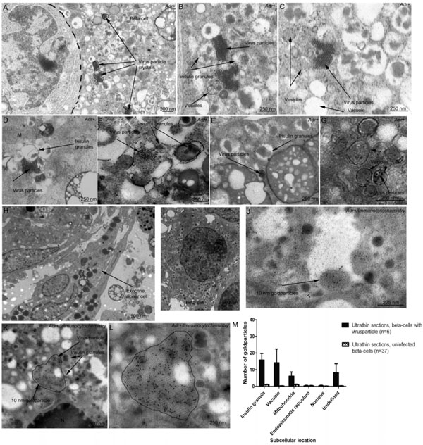
Fig. (5) Ultrastructural micrographs of CVB-5-inoculated human primary pancreatic cell clusters. (A) Virus particle crystals in a beta-cell.
(B-F) Detailed morphology shows association of virus particles to insulin granules and cytoplasmic membranous vesicles and vacuoles. (G)
Vesicular structures enclosing virus particles (H). No virus particles could be observed in the exocrine cells or in (I) mock-infected betacells.
(J-L) Immunocytochemical images of virus-infected beta-cells. The black dots are enterovirus protein 1 (HEVP1) labeled with
secondary antibodies coupled with 10 nm gold particles. (M) Subcellular location of HEVP1 in ultrathin sections of beta-cells with
observable virus crystals (n=6) and mock-infected beta-cells (n=37). The bars represent the mean number of gold particles per beta-cell
section+SEM, not including the gold particles in the large virus crystals. N=Nucleus, M=Mitochondria.